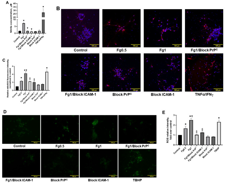
Figure 4.
Fibrinogen (Fg)-induced production of nitrite, mitochondrial superoxide, and the generation of reactive oxygen species (ROS) in neurons. The cells were treated for 2 h with medium alone (control), 0.5 mg/mL of Fg, 1 mg/mL of Fg, 1 mg/mL of Fg in the presence of a cellular prion protein (PrPC)-blocking peptide (Fg1/block PrPC), 1 mg/mL of Fg in the presence of a function-blocking antibody against intercellular adhesion molecule-1 (ICAM-1) (Fg1/block ICAM-1), the prion protein-blocking peptide alone, or the ICAM function-blocking antibody alone. Hirudin was present in all experimental groups to prevent the conversion of Fg to fibrin. (A) The graph depicts the concentration of nitrite measured in media collected from treated neurons using a Griess assay. The experimental group with tumor necrosis factor alpha (TNFα) co-stimulated with interferon gamma (IFNγ) was used as a positive control (TNFα/IFNγ). (B) Representative images show superoxide production by neuronal mitochondria detected via mitoSOX (red). In these experiments, TNFα/IFNγ was used as a positive control as well. Cellular nuclei were labeled with 4′,6-diamidino-2-phenylindole (blue). (C) The summary of the image analyses for the detection of Fg-induced mitochondrial superoxide generation in neurons. (D) Representative images show ROS generation by neurons. Tert-butyl hydroperoxide (TBHP), a common inducer of ROS production, was used as a positive control in this assay. (E) The summary of image analyses for the detection of Fg-induced ROS generation in neurons. p < 0.05 in all; ∗—vs. Control, †—vs. Fg0.5 and ‡—vs. Fg1; n = 4.

