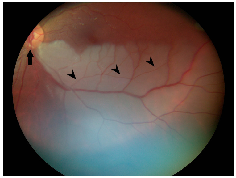
Figure 2.
Color fundus photograph of the left eye at presentation shows retinal whitening of the inferior fundus consistent with diagnosis of branch retinal artery occlusion. Note the yellow refractile body within the inferior arterial arcade at the disc margin (arrow) and attenuation of peripheral arterioles (arrowheads).

