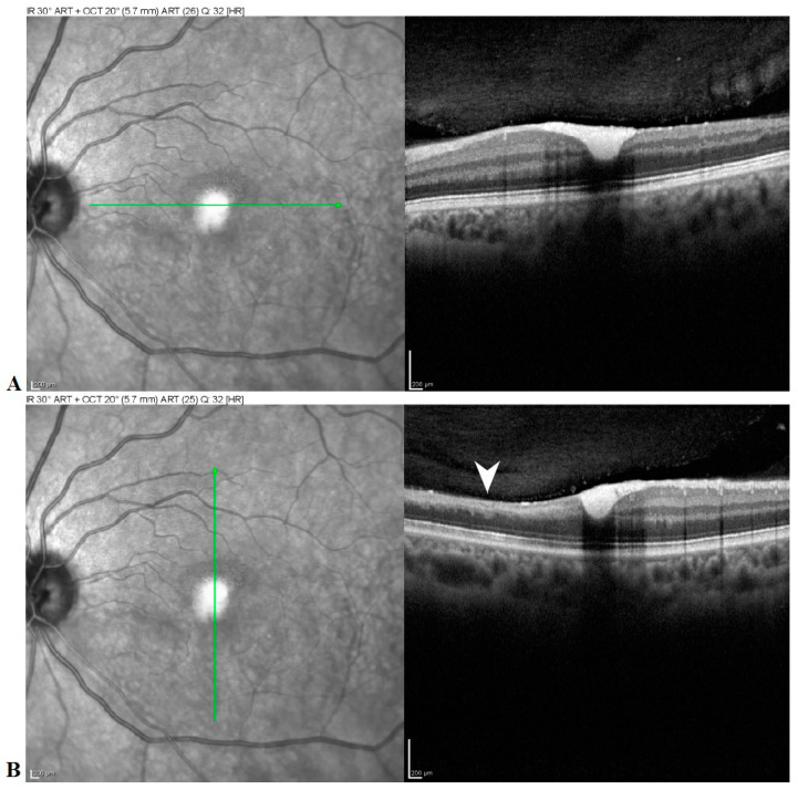
Figure 5.
Horizontal (A) and vertical (B) spectral domain OCT B-scans show hyperreflective material deep to the attachment between the posterior hyaloid and the internal limiting membrane (ILM) and filling the foveal depression suggestive of residual hemorrhage. Note the thinning of the inner retinal layers inferiorly from recent branch retinal artery occlusion (arrowhead).

