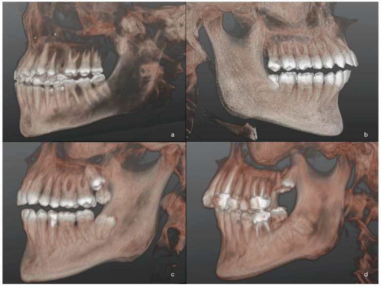
Figure 3.
Three-dimensional images of M3M after 3D reconstructions of CBCT scans in patients of L-GA group. Different levels of M3M position in relation to the adjacent alveolar crest were reported: the most superficial M3M, completely erupted (a); partially impacted M3M in horizontal position (b); completely impacted M3M in the ramus with distoangular position (c); the deepest impacted M3M with roots closer to mandibular canal (d).

