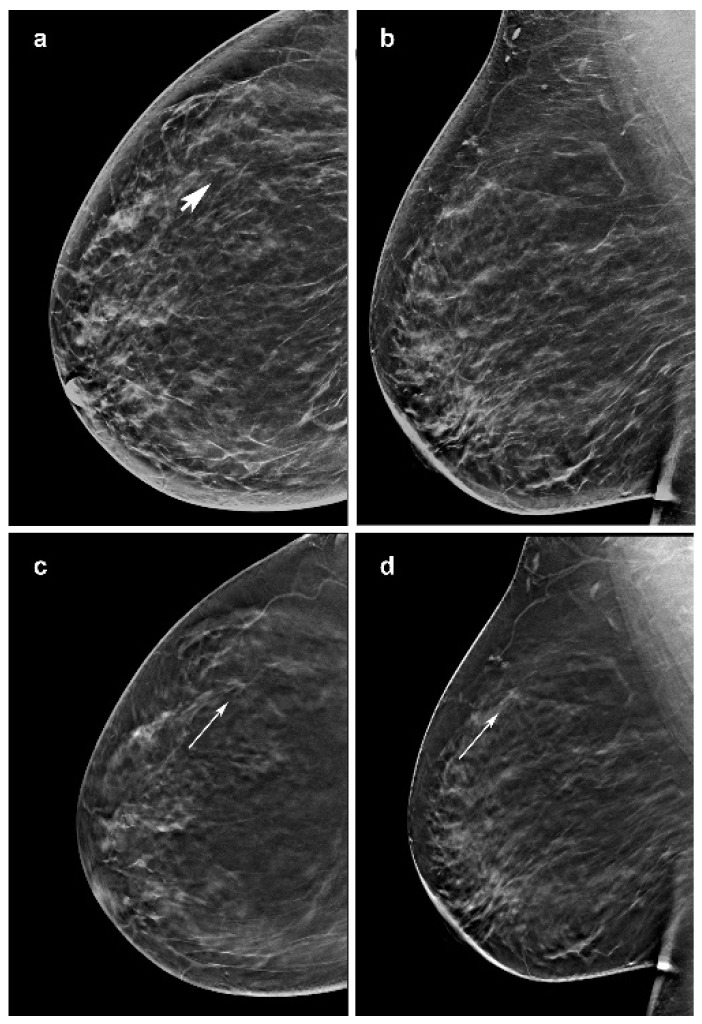
Figure 2.
Invasive lobular carcinoma presenting as a mass in a 57-year-woman, diagnosed at screening. (c,d) Craniocaudal and mediolateral oblique images of the right breast from slices of the DBT portion of the screening study demonstrate a poorly defined 7-mm mass in the outer superior quadrant (arrow). (a) It is less defined in the craniocaudal synthesized mammography (arrow head) and not visible in the mediolateral oblique synthetized mammography oblique image (b).

