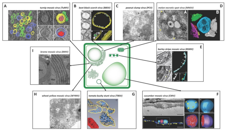
Figure 2.
Structure and origin of plant positive-strand RNA virus replication organelles. (A) 3D architecture of TuMV-induced complex membrane structures. Overview of a single slice of a tomogram of a TuMV-infected vascular parenchymal cell. (upper right) The 3D model shows a SMV with fibrillar material inside and with an adjacent intermediate tubular structure. (lower right) 3D model of a DMV with a core of electron-dense materials. Yellow, SMVs; light red, electron-dense materials; green, intermediate tubular structures; light blue, outer membranes of DMVs; dark blue, inner membranes of DMVs; dark red, the electron-dense materials inside DMVs [4]. (B) Dilated ER of BBSV-infected cells with SMVs (left) and 3D surface reconstruction of the tomogram corresponding to the intact spherules (right) depicting the outer ER membrane (yellow), BBSV-induced spherules (gray), and fibrillar materials inside the spherules (green). Scale bars 100 nm [3]. (C) Electron microscopy of MVB structures in PCV-infected BY-2 protoplasts. White arrows indicate clusters of vesicles. Single arrowheads correspond to MVB; MVB containing disordered membranous vesicles are indicated by black arrowheads, whereas those containing one row of vesicles that are surrounded by a single membrane are indicated by white arrowheads. White asterisks correspond to electron-dense material without detectable vesicles [5]. (D) TEM analysis and 3D reconstruction of MNSV-induced altered mitochondria. (left) TEM image of altered mitochondria. Numerous vesicles were observed on the external surface as well as internal large invaginations and internal dilations (star), or both. Yellow arrowheads indicate the pores connecting the lumen of the dilation to the surrounding cytoplasm. (right) 3D model of MNSV-induced altered mitochondria (blue, yellow, red, and purple) with large dilations inside and close interactions with lipid droplets (grey) and chloroplasts (green) [11]. (E) BSMV-induced chloroplast membrane rearrangement and 3D model of altered chloroplast membranes. (left) Tomogram slices of altered chloroplast membranes from leaves of BSMV-infected N. benthamiana. The arrowheads indicate the same spherules in different slices. (right) 3D model of remodeled chloroplast membranes induced by BSMV indicating the outer chloroplast membrane (cyan), inner chloroplast membrane (gray), and spherules derived from the outer membrane (yellow) [13]. (F) 3D visualization of remodeled tonoplasts in CMV-infected cells. (upper left) Tomogram slice of a CMV-infected N. benthamiana leaf cell. CMV-induced spherules are observed on a vacuolar membrane and in a MVB (arrowheads). The cell wall (CW), cytosol (Cy), and vacuole (Va) are indicated. Scale bar 500 nm. (lower left) 3D model depicting the vacuolar membrane (dark blue), MVBs (light blue), spherules on the vacuolar membrane and in the MVBs (yellow), and a membrane compartment (purple) with virus particles (red). (upper left) 3D model of the MVB with spherules open to the cytosol. (lower left) 3D model of the membrane compartment with virus particles. Scale bars 200 nm [14]. (G) 3D reconstruction of TBSV ROs in wild-type yeast cells characterized by peroxisome-peripheral MVBs depicting the MVB membranes (yellow), vesicle-like spherules (blue) located close to a mitochondrion (red) [9]. (H) Electron micrographs of the mesophyll cells of WYMV-infected wheat. The presence of membranous inclusion body structures in the cytoplasm. The ER, membranous inclusion (MI), mitochondria (Mt), pinwheel inclusion (PW), and virus particles (VP) are labelled [6]. (I) A series of 2–7 appressed layers of double-membrane ER in yeast cells expressing both 2a pol and 1a of BMV, double-membrane ER layers are separated by regular, 50–60-nm spaces, the nucleus (Nuc) and cytoplasm (Cyto) are indicated. Scale bars 100 nm [7] Copyright (2004) National Academy of Sciences, U.S.A. The different parts were reproduced with permission.

