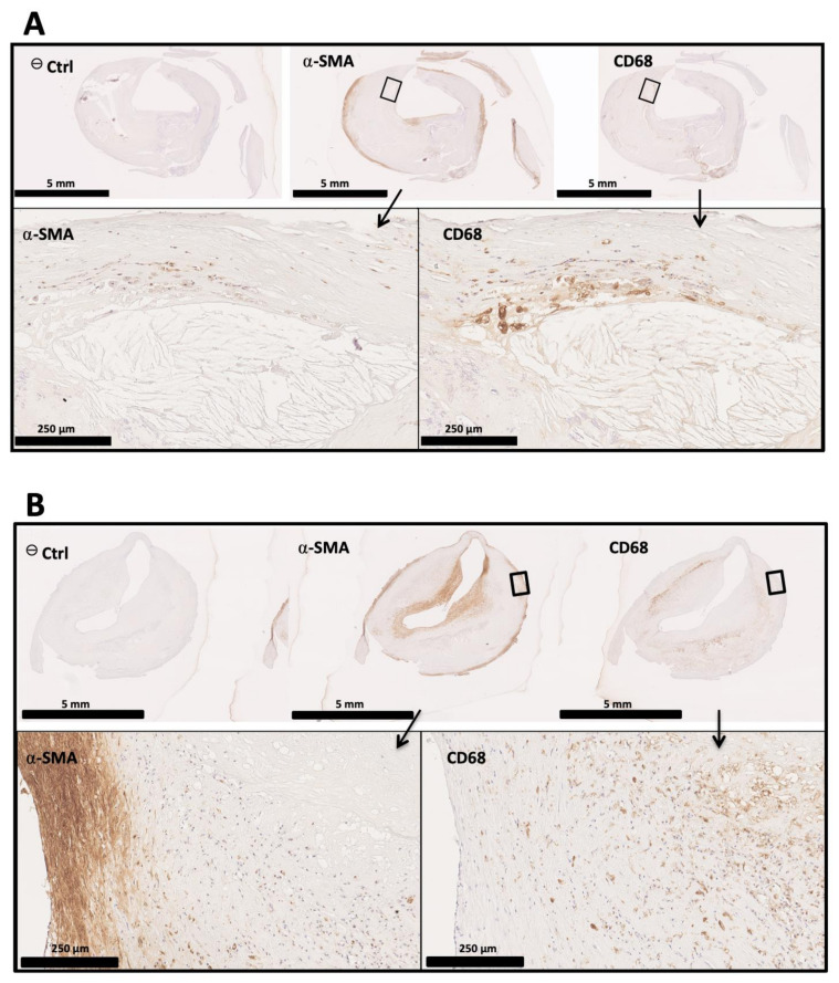
Figure 2.
Immunohistological analysis of human atherosclerostic carotid samples. Human atherosclerotic carotid sections were stained for αSMA and CD68. Higher magnifications show areas positive for both staining, containing cells which seem to express both SMA and CD68, suggesting a phenotypic transition of VSMCs from contractile or synthetic towards a phagocytic subset. (A) Example of an advanced atherosclerotic section with an important necrotic core and a thin fibrous cap (few αSMA-positive cells). Higher magnifications of the delineated area show the interface between the fibrous cap and the underlying necrotic core containing cholesterol crystals. (B) Section of human atheromatous lesion with a thick fibrous cap. Higher magnifications show the interface between the media and the necrotic core, containing cells with potentially intermediate phagocytic/contractile phenotypes.

