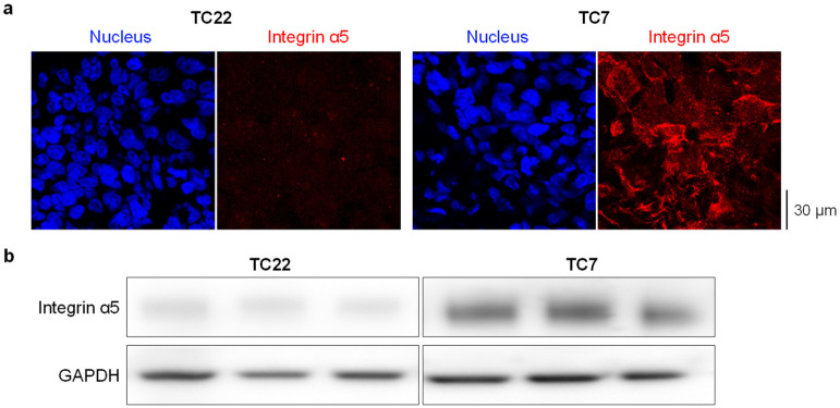
Figure 2.
Immunofluorescence (a) and Western blot (b) on GBM-PDX tumor TC7 and TC22 presenting high and low levels of α5 integrin, respectively. In immunofluorescence, detection of integrin α5 (in red) was realized with AB1928 antibody followed by a secondary antibody coupled to Alexa Fluor® 647. DAPI staining is shown in blue. One representative image per condition is shown (magnification ×63). In Western blot, detection of integrin α5 was realized in 3 xenografts from 3 different mice with H104 antibody. Anti-GAPDH antibody was used as a loading control antibody.

