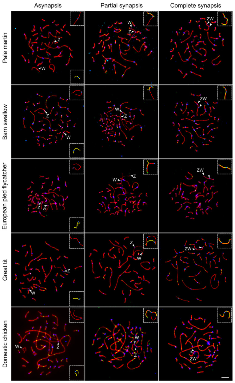
Figure 1.
Synaptic configurations of the ZW SCs during pachytene in five bird species visualized by anti-SYCP3 (red), anti-MLH1 (green) and anticentromere (blue) antibodies. Arrowheads indicate centromeres of Z and W chromosomes. Inserts show schematic representations of Z (red) and W (yellow) SCs. Bar—5 µm.

