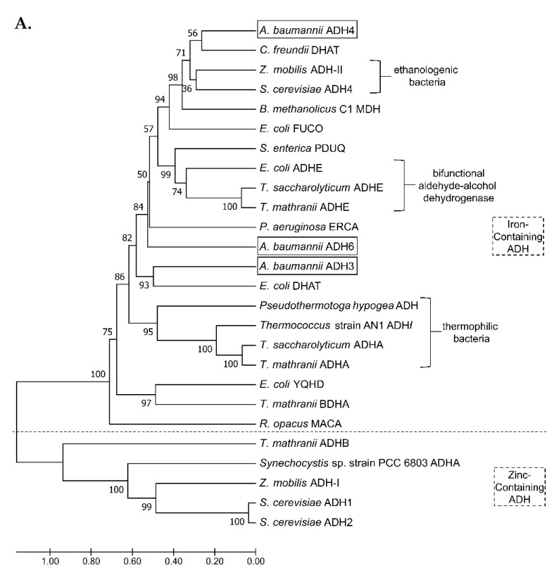
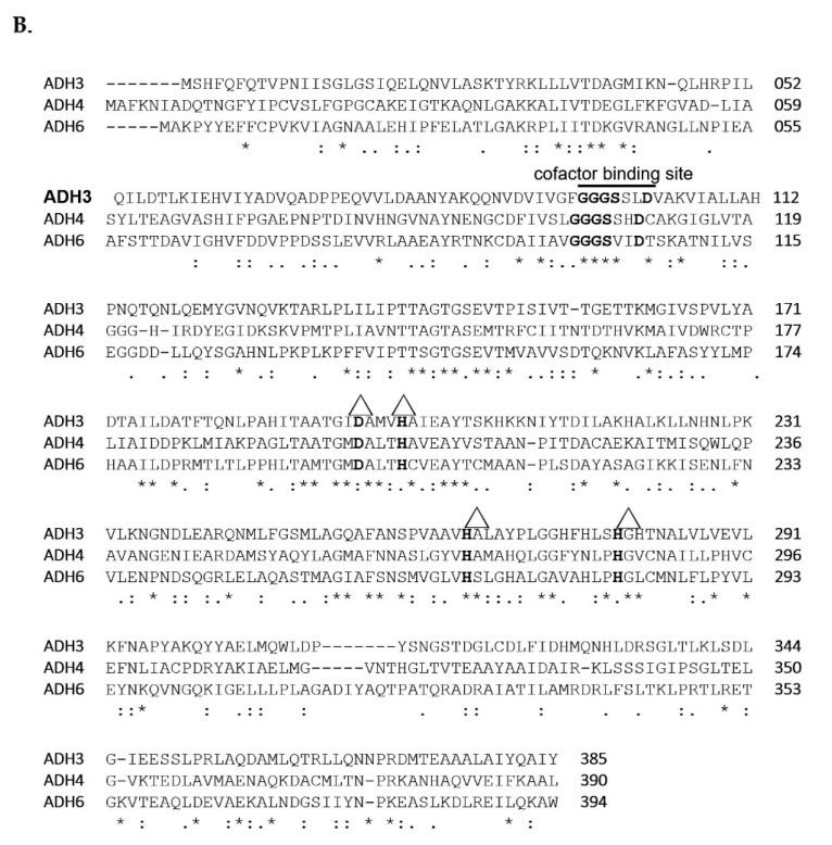
Figure 1.
(A) Cladogram of iron-containing Adh genes in A. baumannii compared with 23 other Adh genes from 14 organisms. The scale bar indicates the number of nucleotide substitutions per site. (B) Amino acid alignment of three FeADHs (ADH3, ADH4, ADH6) in A. baumannii. Δ indicates the positions of the four key iron-binding sites (D, H, H, H). Asterisks (*) indicate positions with single fully conserved amino acid residues; colons (:) indicate positions with conservation between residues of strongly similar properties (scoring > 0.5 in the Gonnet point accepted mutation 250 matrix); and periods (.) indicate positions with conservation between groups of weakly similar properties (scoring ≤ 0.5 in the Gonnet point accepted mutation 250 matrix).


