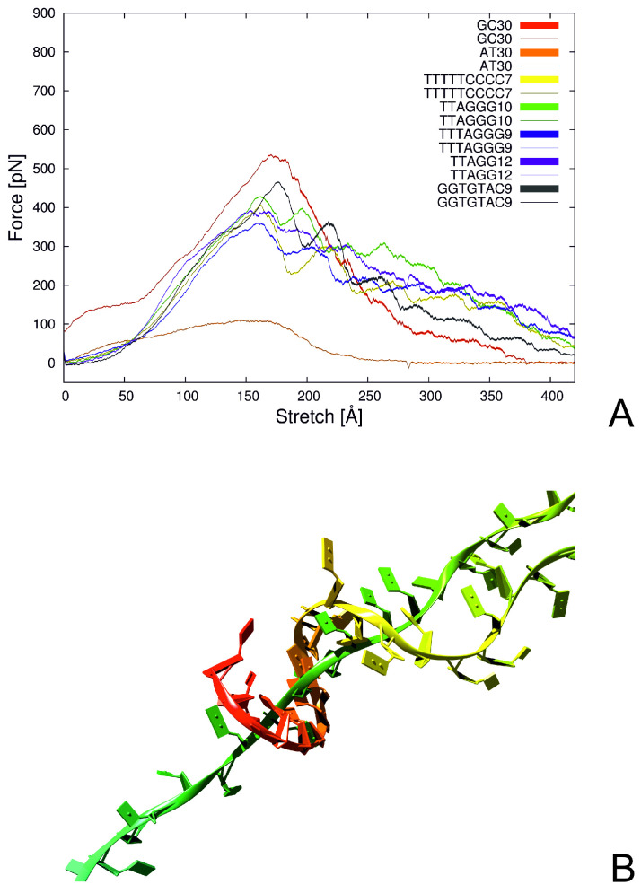
Figure 9.
(A) Plot of the force needed to stretch the DNA perpendicular to the hydrogen bonds between bases, as a function of sequence; Thin lines indicate averages over 64 trajectories and the colored areas indicate one standard deviation. (B) Structure of telomere after regrabbing. The free end (red) wraps around the second DNA chain. Reproduced with permission from A.K. Sieradzan et al., J. Phys. Chem. B, 121, 2207–2219 (2017). Copyright 2015 American Chemical Society.

