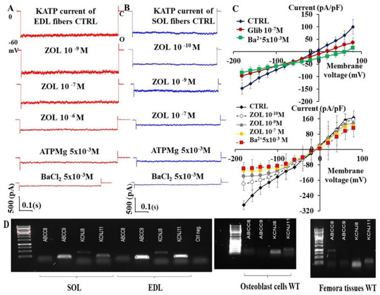
Figure 1.
Effects of increasing concentrations of zoledronic acid (ZOL) on KATP currents recorded in extensor digitorum longus (EDL) (A) and soleus (SOL) (B) muscle fibers and on the current-voltage relationship of primary long bone cells in mice. (A,B) Sample traces from 1 patch per fiber of the KATP currents recorded in excised macro-patches from isolated fibers at −60 mV (Vm) in the presence of 150 mM high K+ ions concentrations on both sides of the membrane patches (O, open current level, C closed current level) in control condition (CTRL) in the absence of added nucleotide or modulators following the excision of the membrane patch from the fibers. The current and the drug action in excised patches was recorded at one voltage close to the resting potential due to the non-voltage-dependent nature of the KATP channel to avoid excessive membrane stress associated with patch isolation. The application of ZOL solution at 10−9 M concentration to the internal side of the membrane patch inhibited the current in SOL fibers by −83% and by −23% in EDL fibers in these patches. The KATP currents were further inhibited by ATPMg 5 × 10⁻³ M applied on the internal side of the macro patches in either fibers. The application of the BaCl2 solution, an unselective KIR blocker, at 5 × 10⁻³ M concentration fully inhibited the residual current, indicating that the ZOL inhibited KATP channel currents in these fibers. (C) Current/voltage relationship of primary long bone cells and effects of KATP channel blockers. The KATP currents were recorded in C-A patches from primary long bone cells in physiological asymmetrical K+ ions condition in the absence of CTRL, presence of glibenclamide (Glib), or increasing concentrations of ZOL solution, followed by BaCl2 solutions applied at the end of protocol period. Glib and ZOL markedly reduced the KATP currents at nanomolar concentrations in primary long bone cells, especially at negative membrane potentials. Each data point was obtained by 5–10 cells. (D) PCR experiments evaluating the expression of the KATP channel genes in Soleus (SOL) and Extensor digitorum longus (EDL) muscles and in femora bone and primary cells from calvaria mice. Abcc9 and Kcnj11 genes were identified in EDL muscle and in SOL muscle. The EDL and SOL muscles also showed the Kcnj8 gene expression. A sample PCR gel shows an intense band at the Kcnj8 gene, suggesting a high expression of KIR6.1 subunit and a low expression of the Kcnj11/KIR6.2 subunit in bone cells. SUR subunits were found. PCR experiments were performed in triplicate.

