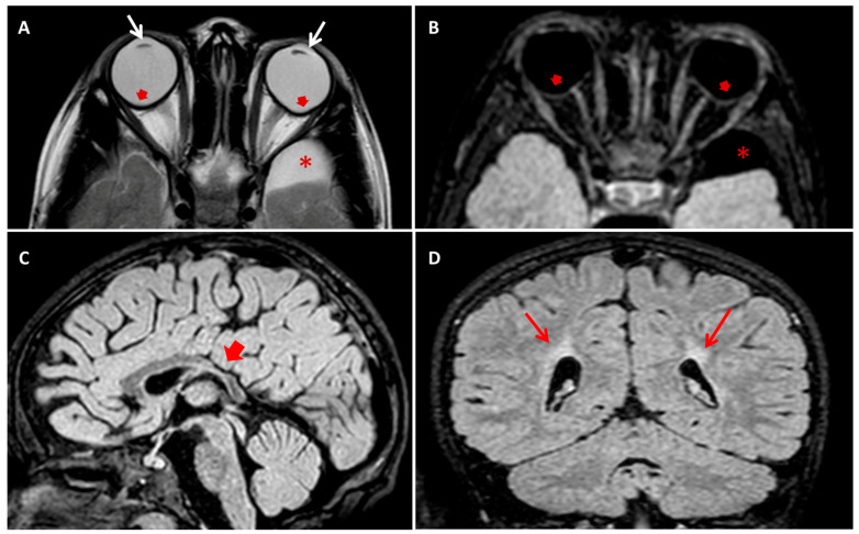
Figure 2.
(A) Axial T2 turbo-spin echo (TSE) and (B) axial T2 fluid attenuated inversion recovery (FLAIR) brain MRI images showing bilateral crystalline lens thinning (white arrows in (A)) and circumscribed outpouching along the posterior aspect of the ocular bulbs, temporal to the optic disc (red arrows in (A,B)). Note a well circumscribed left temporo-polar arachnoid cyst (* in (A,B)). (C) Sagittal T2 FLAIR and (D) Coronal T2 FLAIR brain MRI images showing thinning of posterior trunk of corpus callosum (red short arrow in (C)) and bilateral deep paratrigonal white matter hyperintensity due to periventricular leukoencephalopathy (red arrows in (D)).

