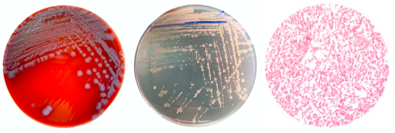
Figure 4.
Wohlfahrtiimonas chitiniclastica colonies on blood agar (Merck KGaA, Germany) and non-selective chromogenic agar (UriSelect, Bio-Rad Laboratories, Hercules, CA, USA) (left, center). Microscopic view of the morphology of W. chitiniclastica (right). Olympus BX40 microscope, Olympus Czech group, s.r.o., magnification 100×. The specimen is stained according to Gram.

