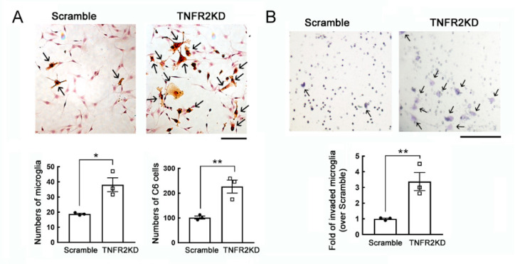
Figure 1.
An increase in the growth and invasive ability of the LPS-primed mouse BV2 microglia cell line with TNFR2 gene knockdown in the co-culture with C6 glioma cells. (A). Scramble-BV2 cells and TNFR2KD-BV2 cells were treated with 10 ng/mL of LPS for 24 h, and then co-cultured with C6 glioma cells for another 24 h. The cultures were subjected to B4 isolectin (IB4) staining to identify BV2 cells. Scramble-BV2 cells or TNFR2KD-BV2 cells with IB4-positive staining show an active hypertrophic shape (arrows). The quantification of IB4+-BV2 cells and C6 glioma cells in the co-cultures were quantified under a microscope. (B). Scramble-BV2 cells and TNFR2KD-BV2 cells were seeded on the upper chamber compartment and primed with 10 ng/mL of LPS for 24 h. The cells were then co-cultured with C6 glioma cells replated onto the lower compartment for 6 h. The invasive TNFR2KD-BV2 cells (arrows) were fixed, stained with crystal violet, and quantified under a microscope. Data are presented as mean ± SEM. The experiments were repeated three times. Each dot (● or □) represents one-time experiment. Scale bar in A, 100 μm; in B, 50 μm. * p < 0.05, ** p < 0.05 versus Scramble.

