Figure 6.
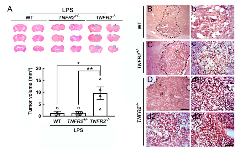
Enhanced tumor growth of mouse GL261 glioma cells in LPS-primed TNFR2 deficient mice. (A) WT, TNR2+/− or TNR2−/− mice were intraperitoneally challenged with LPS at 0.5 mg/kg daily for 7 days, and then the three animal groups were treated with stereotactic intracerebral implantation of mouse GL261 glioma cells. The brain was removed at 14 dpi and sectioned into 20 μm thick cryosections of the three animal groups for H/E staining (upper panel). The bottom panel shows the quantitative data for the tumor volume (mm3), which are presented as mean ± SEM (WT-LPS mice = 4, TNFR2+/−-LPS mice = 5, TNFR2−/−-LPS mice = 4). Each symbol (O, □, or △) represents an animal. * p < 0.05, ** p < 0.01 compared to the WT-LPS mice. (B–D) The brain tissue sections collected from glioma-bearing WT (B,b), TNFR2+/− (C,c), and TNFR2−/− (D,d1–d3) mice with a 7-day LPS injection were subjected to Ki67 immunostaining. Scale bar in A, 5 mm; in (B–D), 200 μm; in (b–d), 50 μm.

