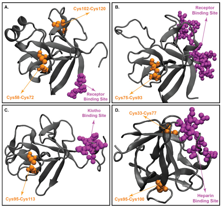
Figure 4.
Crystal structures of endocrine FGFs. (Panel-A): Crystal structure of FGF19. (Panel-B): the crystal structure of FGF21. (Panel-C): the crystal structure of FGF23. (Panel-D): the crystal structure of paracrine FGF2. The protein backbone is represented as a grey cartoon, Disulfide bonds are shown as yellow spheres and receptor binding sequence is represented in magenta; All the crystal structures were prepared using visual molecular dynamics (VMD) software [176].

