Figure 6.
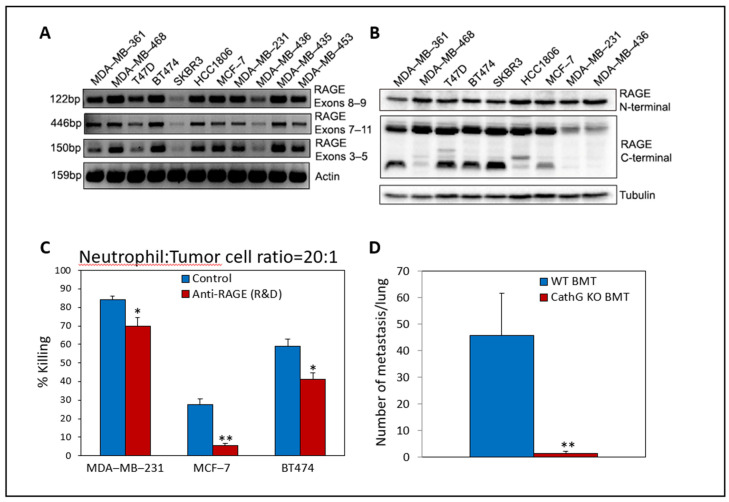
RAGE expression in various human cancer cells and its involvement in neutrophil-mediated cytotoxicity. (A). PCR analysis of mRNA levels as determined by using primer pairs for the indicated RAGE exons and primer pairs for β-actin. (B). Western blot analysis of protein expression levels as determined by using antibodies to the N-terminal part of RAGE (A-9, Santa Cruz, sc-365154), the C-terminal part of RAGE (Abcam, ab3611) or α-Tubulin (Sigma, clone DM1A). (C). Neutralizing antibodies to human RAGE (R&D) (AF1145; 0.5 μg/mL) inhibited neutrophil-induced tumor cell killing toward MCF-7 breast cancer cells and to a lesser extent toward MDA-MB-231 and BT474 breast cancer cells. n = 3 * p < 0.05, ** p < 0.01. (D). AT3 breast cancer cells do not form metastasis in mice, which have been transplanted with Cathepsin G KO bone marrow cells. One hundred thousand GFP-expressing AT3 breast cancer cells were injected intravenously into mice that had been transplanted with either wild-type (WT) or CathG KO bone marrow. The number of GFP-positive metastatic foci in the lungs were counted 8 days later. n = 4 for control and n = 3 for CathG KO. ** p < 0.01. BMT—bone marrow transplanted. These experiments were performed in the laboratory of Prof. Zvi Granot according to the ethics of the Hebrew University’s Institutional Committees.

