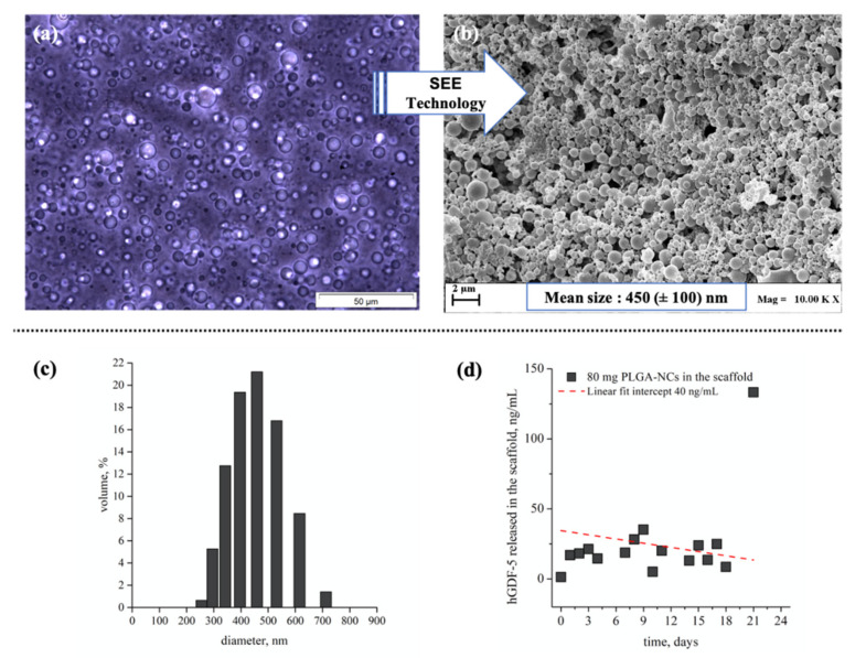
Figure 6.
Images of emulsion and derived PLGA-NCs obtained by SEE technology, particle size distribution, and hGDF-5 release profiles within the 3D environment. Optical microscope image of emulsion (a) and electronic microscope image (b) of carriers obtained after emulsion processing by SEE technology; size distribution data of PLGA carriers expressed as volume percentage (c); in vitro hGDF-5 release profile (ng/mL/day) monitored at 37 °C and 100 rpm by ELISA-based assay from 80 mg of carriers, as loaded in each construct (d); n = 2.

