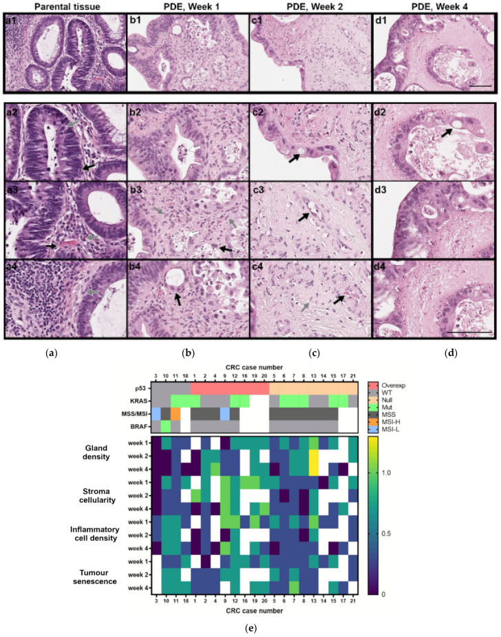
Figure 3.
Histopathological and molecular characterisation of CRC-PDE cultures. (a–d) Representative HE images of the parental tumour and derived explants (PDE) over 4 weeks of culture (for case CRC2). Gland-forming tumour cells (a1–a3,b2,d3), mitotic cells (a2, grey arrow), vascular stroma (a3,c3, black arrows), stromal fibroblast (a4,b3,c4, grey arrows), tumour infiltrating lymphocytes (a2, black arrow) and plasma cells (a3, grey arrow), erythrocytes (c4, black arrow), senescent cells (b3,b4,c2,d2, black arrows), necrotic cells (b2,b3 white arrows), acellular stroma (d4). Scale bars, 100 µm. (e) Heatmap of the histopathological features along culture time (week 1—days 7–9; week 2—days 14–17; week 3: days 25–28) and mutation and protein alterations associated with CRC carcinogenesis, for the 21 CRC cases analysed. Scores for morphological features were performed considering day 0 of culture as the reference and scored as 1 for gland density, stroma cellularity and inflammatory cell density and as 0 for senescence evaluations. White boxes indicate not determined. CRC, colorectal cancer; CRC-PDE, colorectal cancer patient-derived explants; MSS, microsatellite stable; MSI-L, microsatellite instability-low; MSI-H, microsatellite instability-high; Mut, mutated; Overexp, overexpressed; WT, wild type.

