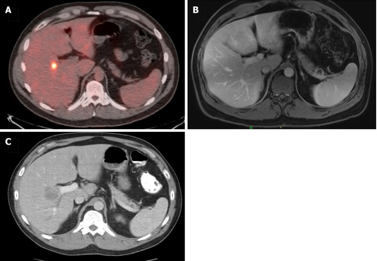
Figure 2.
Disappearing liver metastasis. A: A 36-year-old man presented with a solitary synchronous colorectal liver metastasis to segment 5, seen on staging positron emission tomography-computed tomography scan; B: After 6 cycles of CAPOX plus bevacizumab, followed by pelvic radiation (5040 cGy in 28 fractions), subsequent magnetic resonance imaging (shown) and intraoperative ultrasound were unable to localize the lesion; C: He was then lost to follow-up and returned 18 mo later with an intrahepatic recurrence, seen on surveillance computed tomography scan and confirmed with tissue biopsy.

