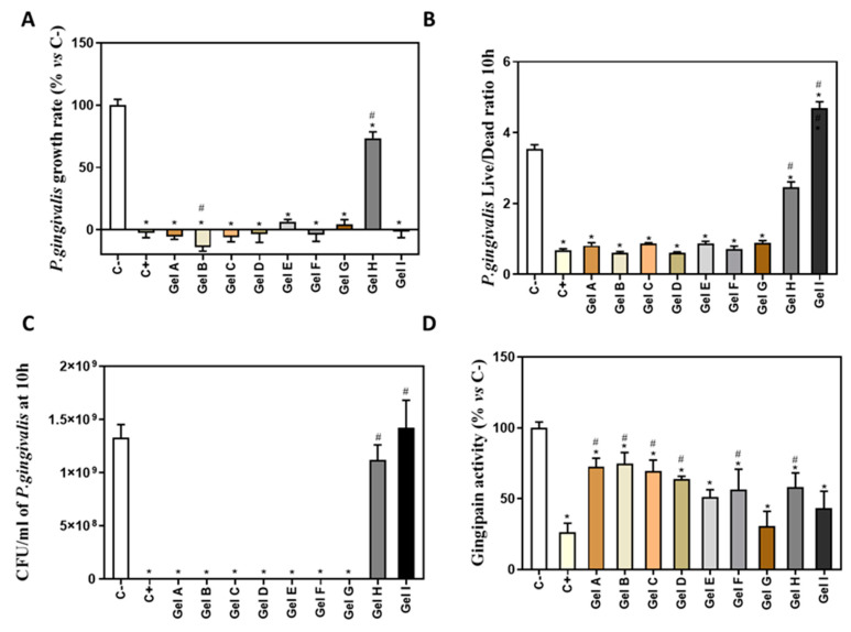
Figure 3.
Antimicrobial activity of different periodontal gels. (A) P. gingivalis growth rate cultured with different periodontal gels (n = 6). (B) P. gingivalis live/dead ratio after treatment for 10 h with different periodontal gels (n = 6). (C) Number of P. gingivalis CFU/mL after 10 h of incubation with the different treatments (n = 4). (D) In vitro gingipain activity from P.gingivalis after 10 h of treatment (n = 6). Results are expressed as % vs. Negative control that was set to 100%. Data represent the mean ± SEM. Negative control (C−) was bacterial suspension without any treatment and positive control (C+) was bacterial suspension with CHX at 0.2%. See Table 1 for the identification of gels used in the study. Results were statistically compared by Kruskal-Wallis for P. gingivalis growth rate and by ANOVA and LSD as post hoc for P. gingivalis live/dead ratio, number of CFU/mL and in vitro gingipain activity: * p < 0.05 treatment vs. negative control. # p < 0.05 treatment vs. positive control.

