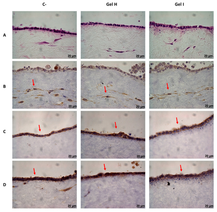
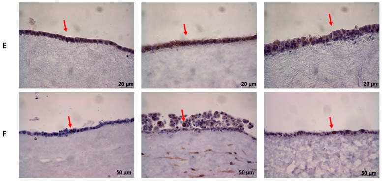
Figure 9.
Histologic characterization of GTE after 72 h of treatment with periodontal gels and LPS stimulation. The images represent an example of (A) H&E staining of GTE; (B) Expression of vimentin (fibroblasts marker); (C) Expression of keratin 19 (epithelial differentiation marker); (D) expression of keratin 17 (epithelial differentiation marker); (E) Expression of involucrin (epithelial differentiation marker); and (F) Expression of Ki-67 (proliferation marker), images were taken at a magnification of 630× (Images of (A–E)) and 400× (Images of (F)). Negative control (C−) was obtained from GTE treated with PBS.


