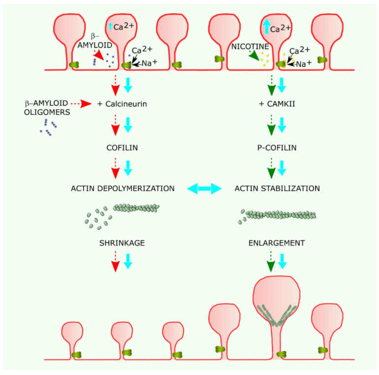
Figure 2.
Possible mechanism of α7 nAChR modulation of dendritic spine morphology and its alteration in Alzheimer disease. The vertical flow diagram on the left shows the pathological cascade, in which β-amyloid oligomers activate α7 nAChR and the calcineurin–cofilin pathway. The increase in intracellular calcium concentration reinforces calcineurin activation. Activated calcineurin dephosphorylates cofilin, which, in turn, depolymerizes actin filaments, leading to spine shrinkage and dysmorphism. Diffusion of cofilin to neighboring spines acts like a chain reaction, spreading the shrinkage along the dendrite. The flow diagram on the right illustrates the protective action of nicotine, which activates α7 nAChRs leading to intracellular calcium increases that activate CAMKII, which, in turn, leads to phosphorylated cofilin (p-cofilin), stabilizing actin filaments. The polymerization and stabilization of actin filaments is associated with spine enlargement.

