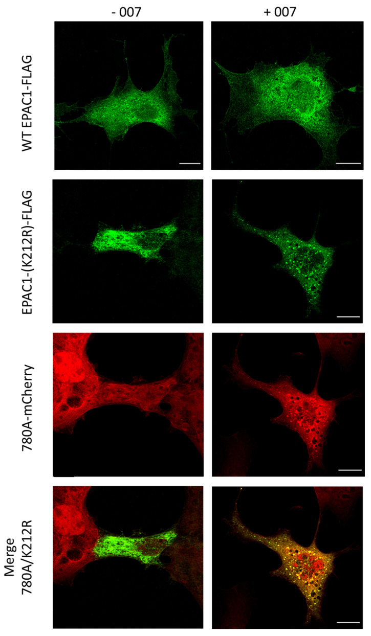
Figure 10.
Localization of Affimer 780A-mCherry with co-transfected wild-type and mutant (K212R) EPAC1 in COS1 cells. COS1 cells were transiently co-transfected with 780A-mCherry and WT-EPAC1-FLAG or EPAC1-(K212R)-FLAG 780A-mCherry and then stimulated ± 100 µM of 007 for 15 min (37 °C, 5% (v/v) CO2). Following fixation, permeabilization and blocking cells were then incubated for one hour each with α-FLAG and goat α-mouse IgG (Alexa Fluor 488 conjugated) primary and secondary antibodies, respectively. Coverslips were then mounted onto glass slides using Prolong™ Glass Antifade Mountant and analyzed on a Leica TCS SP8 STED 3X confocal microscope with a Leica HC PL APO C52 63 × water objective. Samples were excited using a Supercontinuum White laser at 488 and 580 nm for Alexa Fluor 488 and mCherry proteins, respectively, and were detected using a Leica HyD hybrid detector with detection windows of 500–550 nm (Alexa Fluor 488) and 590–650 nm (mCherry). Scale bar: 100 nm.

