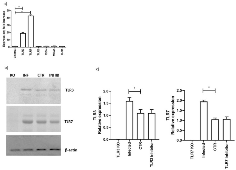
Figure 2.
(a) Calu-3/MRC-5 multicellular spheroids were infected with SARS-CoV-2 at a multiplicity of infection (MOI) of 1.0 for 1 h at 37 °C. Thereafter, the cells were washed and cultured for 48 h. Levels of expression, quantified as fold increase in comparison with uninfected cells of TLR3, TLR7, TLR8, RIG-1, MAD5 and TLR4 are reported and are representative of three independent experiments. (b) Western blot analysis of TLR3, TLR7 and β-actin protein expression in Calu-3/MRC-5 multicellular spheroids (CTR), silenced for TLR3 (TLR3 KO), TLR7 (TLR7 KO) with RNA silencing technology; infected with SARS-CoV-2 with at a multiplicity of infection (MOI) of 1.0 (INF: infected) and treated with TLR3 or TLR7 inhibitors (INHIB: inhibitor). The molecular weights were determined by protein ladder (Bio-Rad, Milan, Italy). Actin was evidenced at 44 kDa, TLR3 and TLR7 at 116 kDa. The images were acquired by Geliance 600 (Perkin Elmer, Milan, Italy). The complete Western blots are reported in Supplementary Figure S2. (c) Evaluation of protein expression by densitometry (GelDoc software; Biorad, Italy), normalized on β-actin content. Data are representative of three independent experiments. Data correspond to the mean +/− standard deviation. * p value < 0.05, calculated with Student’s t-test.

