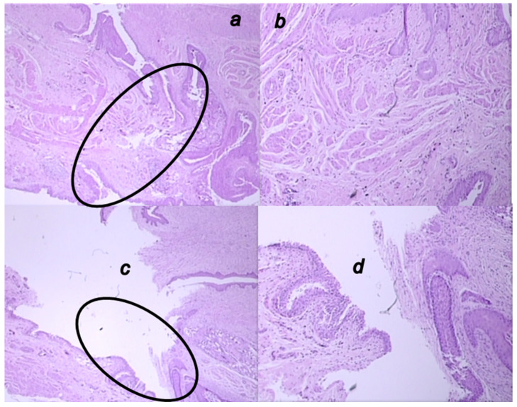
Figure 13.
Microscopic presentation of 6 weeks-vesicovaginal fistula, after the 4 weeks-therapy of fistulous rats in the BPC 157 treated rats (a,b) and control rats (c,d). (a,b) Significant neovascularization with formation of new blood vessels (circle) and mature granulation tissue is visible in the BPC 157-rats (HE; ×20 (a), ×100 (b)). (c,d). Visible open vesicovaginal fistula in control (circle) with moderate inflammation, mature epithelialization, and fully formed subepithelial collagen tissue in the controls (HE; ×20 (c), ×100 (d)).

