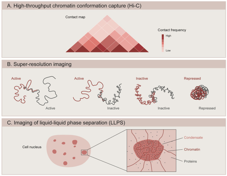
Figure 2.
Methods to study the 3D organization of the genome. (A) High-throughput chromatin conformation capture (Hi-C) generates contact maps that represent the interaction frequency between genomic loci. Studies using Hi-C have revealed that chromatin is organized into topologically associating domains (TADs). (B) Super-resolution microscopy is used to image the spatial organization of different chromatin domains: transcriptionally active (Active), inactive (Inactive), and Polycomb-repressed (Repressed). While active and inactive regions can partially intermix with one another, repressed domains show a more compact configuration and do not overlap with other neighboring domains. (C) Microscopy-based methods are used to assess the ability of chromatin components to form condensates through liquid–liquid phase separation (LLPS). Condensate formation is mediated by the biochemical properties of the macromolecules and their interactions.

