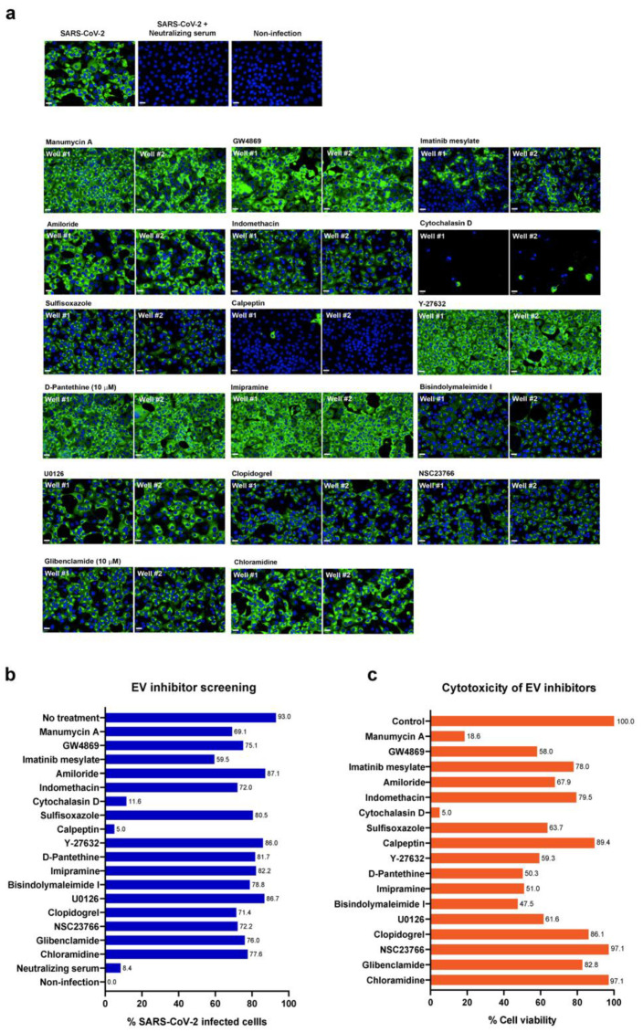
Figure 2.
Screening of 17 known EV inhibitors against SARS-CoV-2 infected cells. Vero E6 cells were infected with SARS-CoV-2 at 25TCID50 for 2 h and subsequently treated with EV inhibitors at post-infection phases for 48 h. Positive convalescent serum of a COVID-19 patient was included as a positive control, and mock infection was performed in parallel as a negative control. The infected cells were then fixed and stained for viral nucleoproteins with anti-SARS-CoV NP mAb. The SARS-CoV-2 infected cells were detected by high-content imaging. (a) The high-content images of the infected Vero E6 cells treated with indicated EV inhibitors at 10 µM are shown. Fluorescent signals: green, anti-SARS-CoV NP mAb; blue, Hoechst. (b) The percentage of the infected Vero E6 was calculated for each condition. The data are presented as an average of two independent experiments. (c) Vero E6 cells were seeded in a 96-well plate overnight and then treated with 10 µM of indicated compounds in a medium for 48 h. Cell viability was examined by the MTT assay. Absorbance was measured at a wavelength of 570 nm. Data were normalized to the solvent control and presented as the percentage of cell viability. Scale bar: 20 µm.

