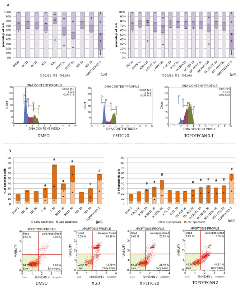
Figure 11.
The impact of X, PEITC, RES, I3C, and their mixtures on cell cycle distribution and induction of apoptosis in HepG2 cells. Panel (A) The percentage of cells in G1/G0, S and G2/M phase analyzed by the flow cytometry after staining with propidium iodide and RNase A. Panel (B) The percentage of cells in the early and late stage of the apoptosis evaluated by the flow cytometry measurements based on fluorescence signal from Annexin V bound to phosphatidylserine externalized in apoptotic cells and a dead cell marker 7-AAD. Topotecan was used as a reference for cell cycle arrest (A) and pro-apoptotic activity (B). Exemplary plots are presented. Results were calculated from three separate experiments (mean ± SEM). (*) above the bar indicates statistically significant differences from the control group, while hashes (#) indicate statistically significant changes in the percentage of total apoptotic cells, p < 0.05.

