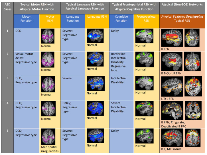
Figure 1.
Comparison of clinical and rs-fMRI findings in ASD patients. Columns 1–3: ASD patient typical motor, language, and frontoparietal network images and interpretation with corresponding discordant phenotypic clinical impairments. Column 4: ASD participant atypical (aberrant) network (non-SOZ, overlapping typical RSN) images. B, bilateral; DCD, developmental coordination disorder; F, frontal; FPN, frontoparietal network; L, left; MT, mesial temporal; Opr, operculum; R, right; RSN, resting state network; vmPFC, ventromedial prefrontal cortex.

