Figure 2.
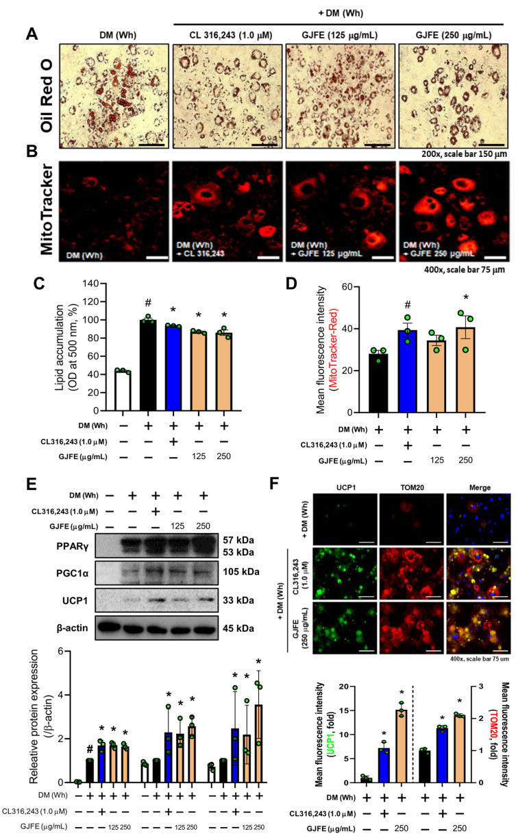
Effect of GJFE on beige trans-differentiation of white-induced 3T3-L1 cells. (A) Intracellular lipid droplets were stained with Oil Red O (magnification 400×, scale bar 75 μm), and (C) the absorbance was detected at 500 nm in white-induced 3T3-L1 cells treated with GJFE (125 and 250 µg/mL) or CL316,243 (1.0 µM) (β3-AR agonist). (B,D) Mitochondrial abundance was obtained by MitoTracker Red staining (magnification 400×, scale bar 75 μm). (E) Protein levels of PPARγ, PGC1α, and UCP1 were analyzed by Western blot analysis. (F) The expression of UCP1 (green), TOM20 (red), and DAPI (blue) were detected by immunofluorescence staining (magnification 400×, scale bar 75 μm). β-actin was used as a loading control for Western blot analysis. All data are expressed as the mean ± SEM of the data from three or more separate experiments. # p < 0.05 vs. DM (Wh)-untreated 3T3-L1 cells, * p < 0.05 vs. DM (Wh)-treated 3T3-L1 cells. GJFE, Gardenia jasminoides fruit extract.

