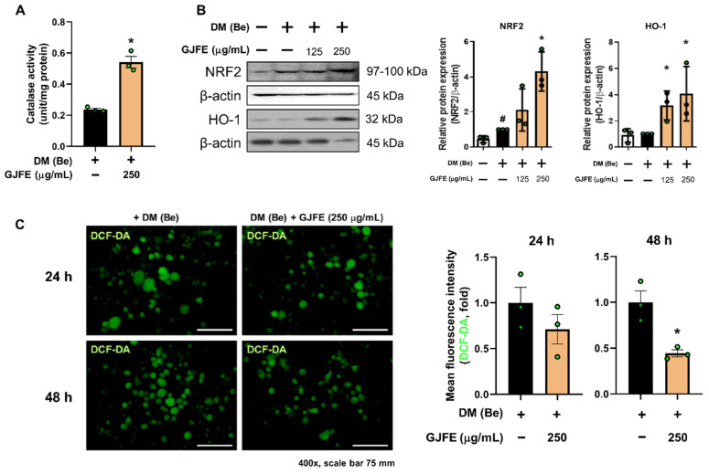
Figure 6.
Effect of GJFE on oxidative stress in beige-induced 3T3-L1 cells. (A) Catalase activity was measured in beige-induced 3T3-L1 cells treated with GJFE (250 µg/mL). (B) Protein levels of NRF2 and HO-1 were analyzed by Western blot analysis. (C) DCF-DA staining was performed at 24 h and 48 h post GJFE treatment in beige-induced 3T3-L1 cells (magnification 400×, scale bar 75 μm). β-actin was used as a loading control for Western blot analysis. All data are expressed as the mean ± SEM of the data from three or more separate experiments. # p < 0.05 vs. DM (Be)-untreated 3T3-L1 cells, * p < 0.05 vs. DM (Be)-treated 3T3-L1 cells. GJFE, Gardenia jasminoides fruit extract.

