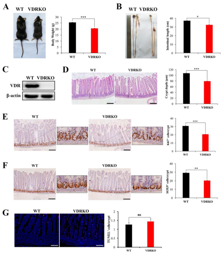
Figure 3.
VDR deficiency impairs the intestinal physiological structure and crypt stem/progenitor cell proliferation. (A) Appearance of wild type (WT) and VDR knockout (VDRKO) mice and quantification of body weights at the age of 8 weeks. n = 12 in each group. (B) Appearance of intestines of WT and VDRKO mice and quantification of intestine length. n = 7 in each group. (C) VDR was evaluated in intestine tissues of mice by WB. (D) Representative H&E images of intestine of WT and VDRKO mice and quantification of crypt depth. n = 6 in each group. Scale bar, 200 μm. (E) Representative IHC images of Ki67 staining of WT and VDRKO mice and quantification of number of Ki67+ cells/crypt. Scale bar, 200 μm. n = 6 in each group. (F) Representative IHC images of SOX9 staining of mice and quantification of number of SOX9+ cells/crypt. n = 6 in each group. Scale bar, 200 μm. (G) Representative IF images of TUNEL staining of WT and VDRKO mice and quantification of TUNEL+ cells/crypt. n = 6 in each group. Scale bar, 100 μm. Values are means ± SD. * p < 0.05, ** p < 0.01, *** p < 0.001.

