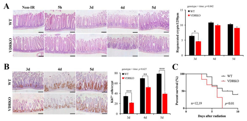
Figure 4.
VDR deficiency suppresses intestinal epithelial regeneration and survival time of mice following IR. (A) Representative H&E images of intestine of WT and VDRKO mice at different times of 0, 5 h, 3 d (days), 4 d and 5 d after 12 Gy IR. Quantitative analysis of the number of regenerated crypts/1250 μm at different times after IR. n = 6 in each group. (B) Representative IHC staining of the expression of Ki67 of WT and VDRKO mice and quantification of Ki67+ cells/crypt at different times after IR. n = 6 in each group. Values are means ± SD. * p < 0.05, ** p < 0.01, *** p < 0.001. Scale bar, 200 μm. (C) Kaplan–Meier survival analysis of mice subjected to 12 Gy IR. The statistical analysis was performed by log-rank test. n ≥ 12 in each group.

