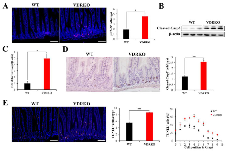
Figure 5.
VDR deficiency aggravates IR-induced DNA damage and crypt stem/progenitor cell apoptosis. (A) Representative IF images showing the γH2AX expression at 5 h post 12 Gy IR and quantification of γH2AX+ cells in crypts. n = 6 in each group. Scale bar, 100 μm. (B) Cleaved Casp3 was evaluated in intestine tissues of mice at 5 h after IR by WB. (C) The Cleaved Casp3 expression was quantitatively analyzed. (D) Representative IHC images of Cleaved Casp3 at 5 h after 12 Gy IR and quantification of Cleaved Casp3+ cells in crypts. n = 6 in each group. Scale bar, 50 μm. (E) Representative IF images of TUNEL staining of WT and VDRKO mice at 5 h after 12 Gy IR. Quantification of TUNEL+ cells/crypt and their positions in crypts. Scale bar, 100 μm. n = 6 in each group. Values are means ± SD. * p < 0.05, ** p < 0.01.

