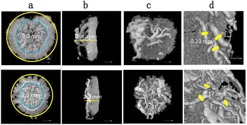
Figure 2.
Representative 3D vascular castings of HO-1 Het (top panel) and WT (bottom panel) placentas harvested at E16.5 using MicroCT. Images show differences in: (a) circumferences; (b) thicknesses; (c) maternal vasculatures; and (d) spiral artery diameters. Reprinted with permission from Ref. [129]. Copyright 2011 Zhao H, et al.

