Figure 2.
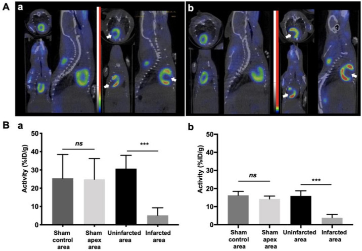
Myocardial infarction resulted in myocardial viability defect evaluated by 18F-FDG microPET imaging. (A) 18F-FDG microPET imaging showed a signal decrease in the infarcted myocardium identified by a white arrow at day 16 (a) and at day 30 (b) post-surgery. (B) Semi-quantitative analysis expressed as percentage of injected dose per gram of mouse (%ID/g mean ± SD) for day 16 (a) to day 30 (b) post-surgery showed a significant decreased in Apex area compared to control area (Meanapex = 5.19 ± 4.10%ID/g, Meancontrol = 30.7 ± 7.25%ID/g, n = 7; *** p = 0.0003 at day 16) and (Meanapex = 3.84 ± 1.81%ID/g, Meancontrol = 15.9 ± 2.81%ID/g, n = 7; *** p = 0.0003 at day 30). In contrast, uptake was homogeneous in sham hearts for both experiment time (Meanapex = 24.8 ± 11.3%ID/g, Meancontrol = 25.4 ± 12.9%ID/g, n = 4; ns: p = 0.443 at day 16) and (Meanapex, 14.3 ± 1.54%ID/g, Meancontrol 16.2 ± 2.20%ID/g, n = 4; ns: p = 0.885 at day 30).

