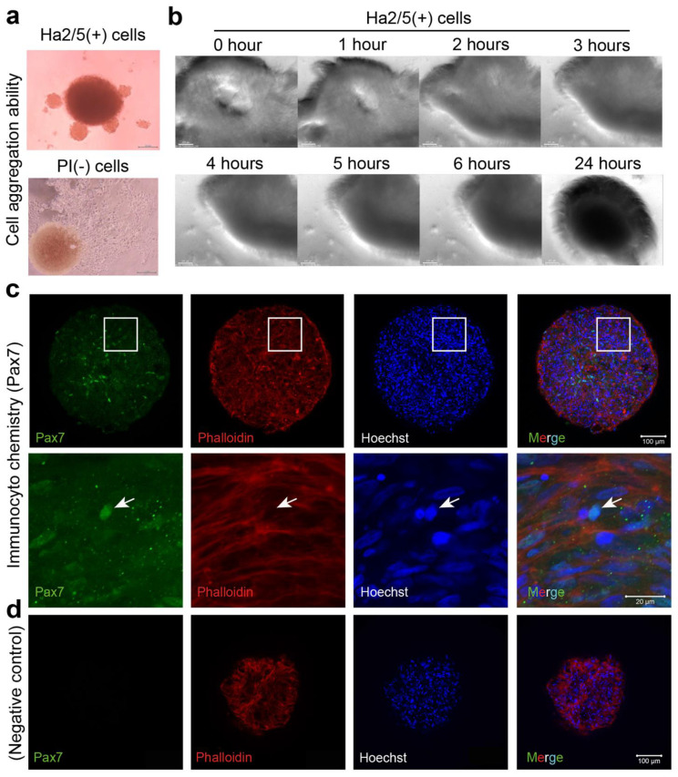
Figure 3.
An analysis of the spheroid-forming capacity of Ha2/5+ cells. (a) Ha2/5 cells (1 × 10⁵) were seeded in 96-well round-bottom plates, centrifuged at 400× g, and then cultured for 24 h. Ha2/5+ cells and unpurified cells (all living cells) were seeded, and photomicrographs taken one week later are shown. The scale bar represents 200 μm. The brown mass (in PI-negative cells) was hematopoietic cells. (b) Images of spheroid-forming cells at 0, 1, 2, 3, 4, 5, 6, and 24 h after seeding the cultured Ha2/5 cells. (c) Analysis of Ha2/5 spheroids by immunostaining. Muscle marker: Pax7 are shown in green. Cyto-skeletal marker (Phalloidin, red) and cell nuclei (Hoechst, blue) are shown. The white square is enlarged on the lower panel. Allowhead indicate the localization of Pax7. (d) Negative controls in Pax7 staining were displayed. The scale bar represents 100 μm.

