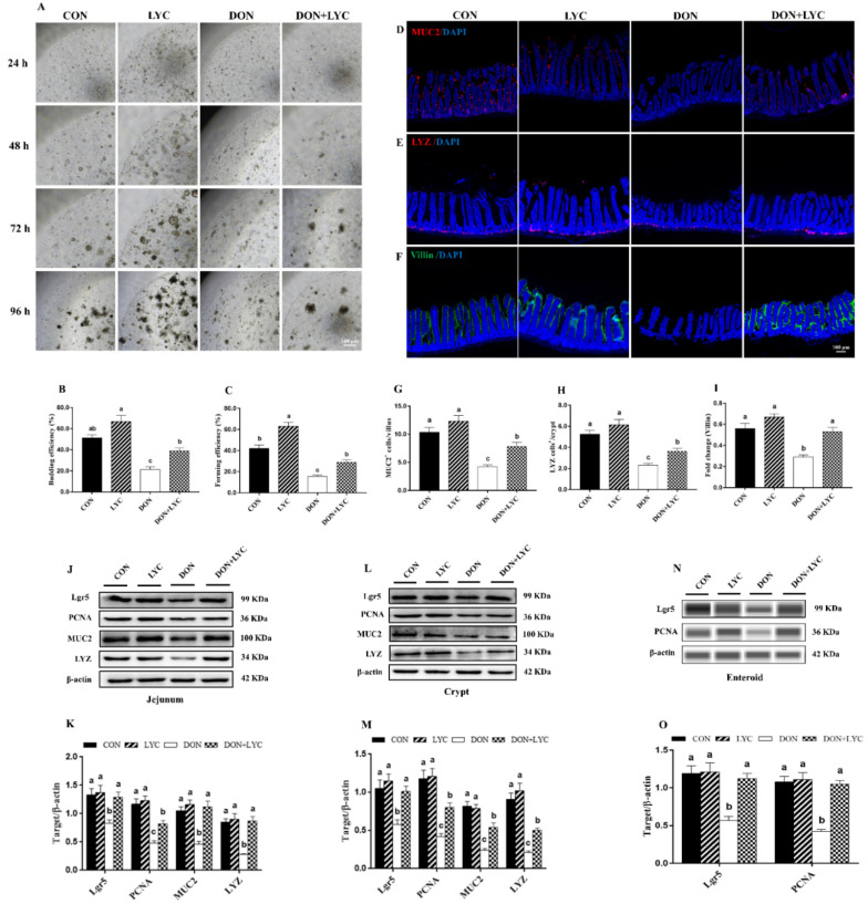
Figure 4.
LYC treatment stabilized the functions of intestinal epithelial cells under DON exposure. (A) Represented images of enteroids expended from crypt stem cells. (B,C) Enteroid forming and budding efficiency. (D–F) Represented images of immunohistochemistry staining with MUC2, LYZ, and Villin antibodies in the jejunum. (G,H) Statistical analysis of MUC2+ cells and LYZ+ cells. (I) Statistical analysis of fluorescence intensity of Villin. (J,K) Protein expression of PCNA, Lgr5, MUC2, and LYZ in the jejunum. (L,M) Protein expression of PCNA, Lgr5, MUC2, and LYZ in the crypt. (N,O) Protein expression of Lgr5 and PCNA in the enteroids of mice. Results are presented as mean ± SEM (n = 3). Columns with different superscripts letters indicating significant difference (p < 0.05).

