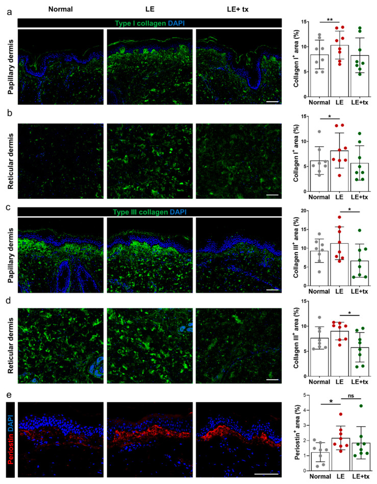
Figure 4.
Treatment with QBX258 decreases type III collagen deposition in lymphedematous tissues. Fibrosis after QBX258 treatment. (a,b) Representative immunofluorescent staining (left panels) and quantification (right panel) in normal, LE, LE + tx biopsy specimens for DAPI (blue) and type I collagen (green) in the papillary (a) and reticular (b) dermis. (c,d) Representative immunofluorescent staining (left panels) and quantification (right panel) in normal, LE, LE + tx biopsy specimens for DAPI (blue) and type III collagen (green) in the papillary (c) and reticular (d) dermis. (e) Representative immunofluorescent staining (left panels) and quantification (right panel) of normal, LE, LE + tx biopsy specimens for DAPI (blue) and periostin (red). Scale bar: 50 μm; * p < 0.05; ** p < 0.01.

