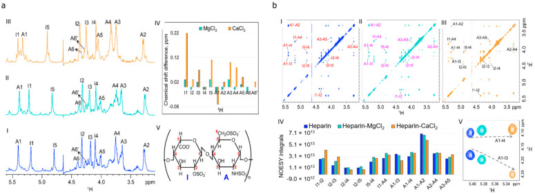
Figure 2.
NMR analyses of magnesium chloride modified heparin: (a) Comparison of a portion of 1D 1H NMR spectra of heparin (I), heparin–Mg2+ (II), and heparin–Ca2+ (III) acquired in 99.95% D2O at 37 °C. Proton chemical shift assignment of iduronic acid, and glucosamine, residues are shown on top of each resonance. (IV) Bar plot showing 1H chemical shift perturbation of heparin in the presence of Mg2+ and Ca2+. 1H chemical shift difference between heparin–M2+ (M = Mg, Ca) and heparin are presented on the y-axis. Positive value indicates the deshielding of respective protons in the presence of M2+ (M = Mg, Ca). (V) Structure of the repeating disaccharide (iduronic acid-glucosamine) unit of heparin; (b) A portion of 2D NOESY spectra of heparin (I), heparin–Mg2+ (II), and heparin–Ca2+ (III) in 99.95% D2O at 37 °C. Assignments are shown for the characteristic NOE cross-peaks. (IV) Bar plot representation of intra- and inter-residue NOE cross peak volumes. (V) Change in NOESY cross peak intensities of inter-residue A1-I4 and A1-I3 protons of heparin (blue), heparin–Mg2+ (cyan), and heparin–Ca2+ (orange).

