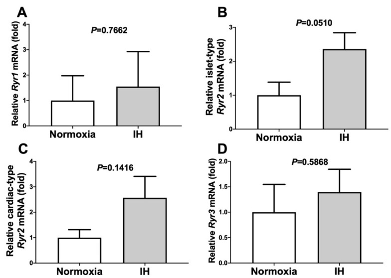
Figure 7.
The mRNA levels of Ryr1 (A), islet-type Ryr2 (B), cardiac-type Ryr2 (C), and Ryr3 (D) in the As4.1 JG cells subjected to normoxia or IH for 24 h. In all cases, the mRNA levels were not elevated by IH (P = 0.7662, P = 0.0510, P = 0.1416, and P = 0.5868 in the Ryr1, islet-type Ryr2, cardiac-type Ryr2, and Ryr3, respectively). The data are expressed as the mean ± SE for each group of six independent experiments (n = 6). The statistical analyses were performed using a Student’s t-test.

