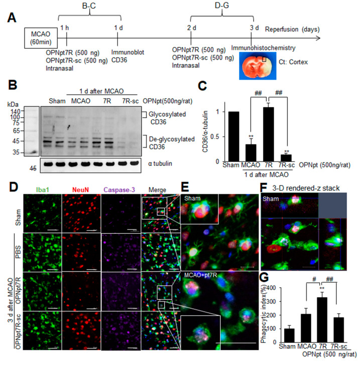
Figure 6.
Enhanced phagocytic activity of microglia by OPNpt7R in the post-MCAO brain; (A) OPNpt7R (500 ng) or OPNpt7R-sc (500 ng) was administered intranasally at 1 h post-MCAO (B,C) or at 2 d post-MCAO (D–G). (B,C) Protein samples were prepared from the indicated region in A at 1 d post-MCAO, and the CD36 level was examined by immunoblot analysis. Representative images are presented (B) and quantified results are presented as the mean ± SEM (n = 3) (C). (D–G) Coronal brain sections were prepared from sham, MCAO, MCAO+OPNpt7R, and MCAO+OPNpt7R-sc groups at 3 d after MCAO and processed for immunostaining with anti-Iba1, anti-NeuN, and anti-activated caspase 3 antibodies. Representative pictures are presented (D–F) and phagocytic index (the ratio of Caspase 3-encapsulated in Iba1 over Caspase 3 in regions (0.2 mm2) indicated as black box in A are presented as the mean ± SEM (n = 10, 10 brain slices from three animals) (G). The images in E are high-magnification photographs of images in the Sham and MCAO+OPNpt7R group (indicated as white boxes). Scale bars represent 50 μm. Sham, sham-operated rats; MCAO, PBS-treated MCAO controls; MCAO+OPNpt7R, OPNpt7R-treated MCAO rats; MCAO+OPNpt7R-sc, OPNpt7R-sc-treated MCAO rats. ** p < 0.01 vs. PBS-treated MCAO controls, and # p < 0.05, ## p < 0.01 between indicated groups.

