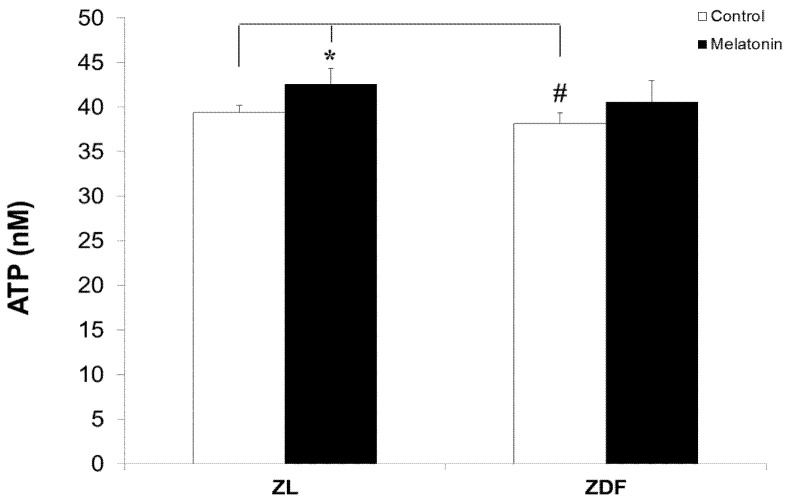
Figure 4.
ATP levels of BAT-isolated mitochondria of different treated groups. Data were normalized for variations in mitochondrial content in an equivalent amount of tissue (micrograms of ATP/mg of tissue wet weight), adjusted to 0.5 mg/mL protein concentration, and expressed as nmol/mg protein. Data are shown as mean ± SD. Superscript characters show significant differences determined by two-way ANOVA followed by the Tukey post hoc test (* p < 0.01, M-ZL compared with C-ZL rats; # p < 0.05, C-ZDF compared with C-ZL rats).

