Figure 2.
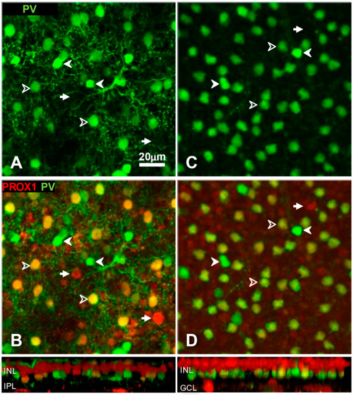
Typing of amacrine cells based on double labelling with parvalbumin (green channel) and Prox1 (red channel) immunoreactivity in cat (A,B) and rat (C,D) retina. Microscopic images show horizontal views of whole-mount preparations at the level of amacrine cells. Side-view reconstructions of representative sub-volumes spanning the INL (top) IPL and ganglion cell layer (GCL) are shown below (B,D). (A,C) Anti-parvalbumin antibody labelled two populations of amacrine cells in both species. The weakly parvalbumin-positive neurons (open arrowheads) include (in cats, A) or are exclusively (in rats C) AII amacrine cells. B, D. The combination of parvalbumin and Prox1 immunolabels reveals three amacrine cell types in both species. Amacrine cells with a strong PV expression are Prox1-negative. Weakly PV-positive amacrine cells are Prox-1 positive. A small population of Prox1-positive amacrine cells is PV-negative (arrows). Scale, 20 μm.

