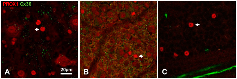
Figure 4.
Prox1 immunoreactive cell nuclei (red channel) in the ganglion cell layer of flat mounted cat (A), rat (B) and mouse (C) retina. Smaller nuclei (arrows) are putative displaced amacrine cells (see details in the text). In the cat retina (A), some ganglion cell somata are also Prox1-immunoreactive. Connexin-36 puncta appear in irregular patches where the neuropil of the inner plexiform layer intrudes between cell bodies, but no somatic plaques can be observed on the Prox1-positive cells. Non-specific labelling by the anti-Cx36 antibody is seen in a bundle of optic fibers in the mouse retina (C). Scale bar 20 μm.

