Figure 5.
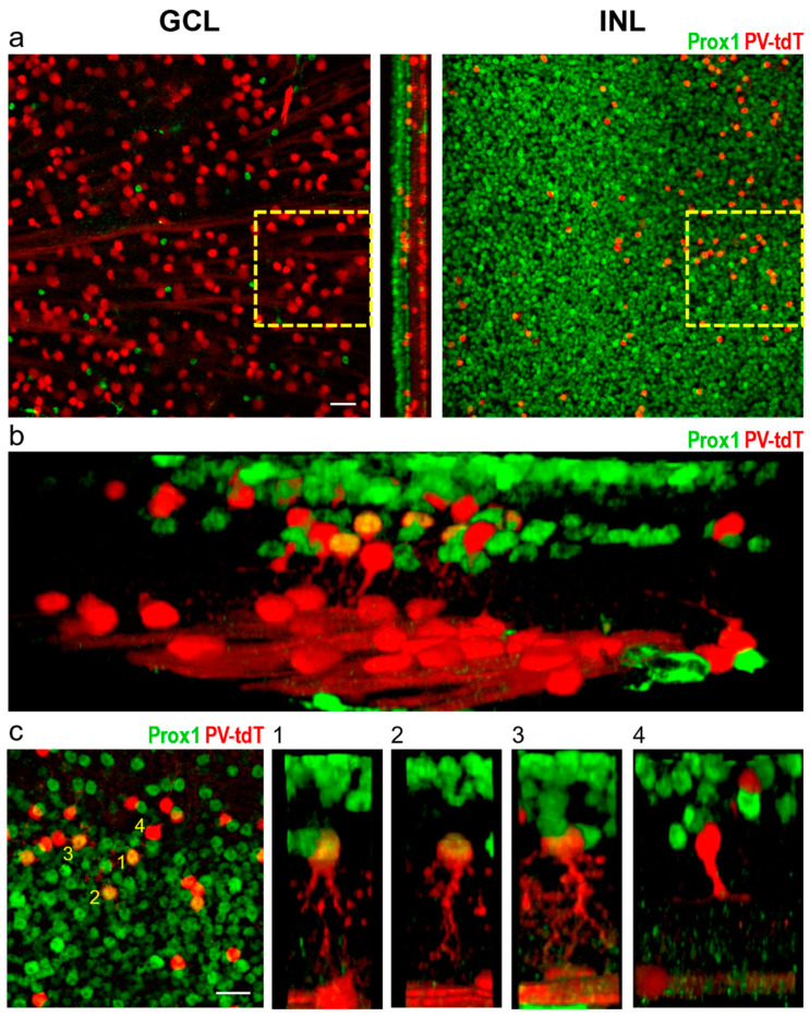
Colocalization of tdT and Prox1 in whole-mount preparation of the PV-tdT mouse retina. Overview images (a) show no colocalization of Prox1 with tdT in the ganglion cell layer (GCL), but in the INL, patches of amacrine cell bodies were double-labelled. The box outlined in yellow broken lines is shown at higher magnification in c. Three-dimensionally rendered and rotated views (b,c) of double labelled cells revealed their typical AII-like dendritic morphology (c(1–3)). Prox1-negative amacrine cells showed sparser, monostratified dendritic arbours reminiscent of wide-field amacrine cells (c(4)). Numbers in the left panel of c identify cells shown on side-views numbered 1 through 4, Scale = 25 μm.

