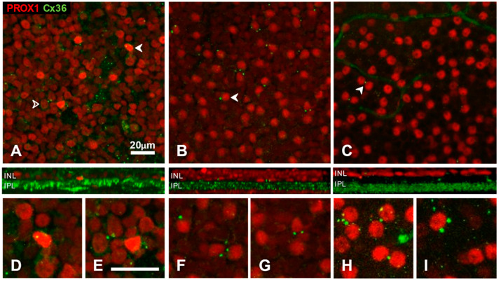
Figure 6.
Somatic Cx36 plaques on Prox1 immunoreactive amacrine cells in whole-mount preparations of cat (A,D,E), rat (B,F,G) and mouse (C,H,I) retinas. Red channel, Prox1; green channel, Cx36. Side-view reconstructions of representative sub-volumes spanning the proximal INL (top) and IPL are shown below (A–C). Somatic Cx36 plaques were found in close apposition to a subset of Prox1-immunoreactive cell bodies. Examples are marked by arrowheads or shown at higher magnification in (D–I). In the cat retina (A), somatic plaques occurred in apposition to both strongly Prox1 positive (solid arrowheads) and lightly Prox1-positive (open arrowhead) cells.

