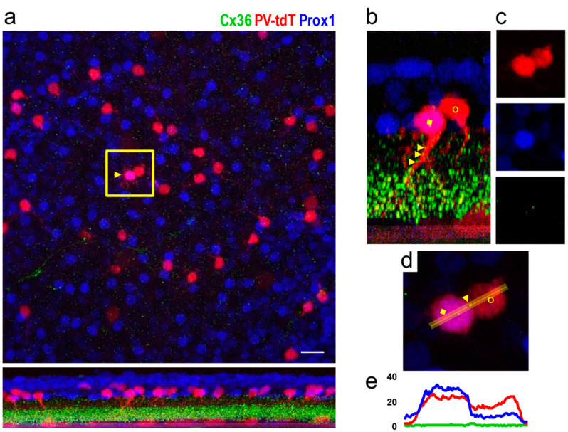
Figure 7.
Colocalization of tdT, Prox1 and Cx36 immunofluorescence in amacrine cell somata of PV-tdT mouse retina shown in a tangential view at the level of the amacrine cell bodies (top of panel (a)) and as an orthogonal section along the line indicated by the yellow arrowhead (bottom of panel (a)). The region enclosed by the yellow box contains an AII amacrine cell (yellow arrowhead, magenta color due to double labelling with Prox1 and tdT) along with a tdT-positive neighbouring non-AII amacrine cell (red). The corresponding dendritic morphology of these cells is readily seen on the side-view in (b) (yellow square, AII amacrine cell; yellow circle, non-AII amacrine cell). Panel (c) shows the boxed region from a with the three channels separated (PV-tdT, Prox1 and Cx36 from top to bottom). Note the dot on the green channel, which indicates a Cx36 plaque where the two cell bodies touch each other. A close-up of this region (d) and an intensity profile, measured across the Cx36 plaque (e) confirm the close apposition of the plaque to both cell bodies. Some Cx36-puncta are also present on the proximal dendrite of the AII cell (b, yellow arrowheads). Markers as in (b). Scale, 25 μm.

